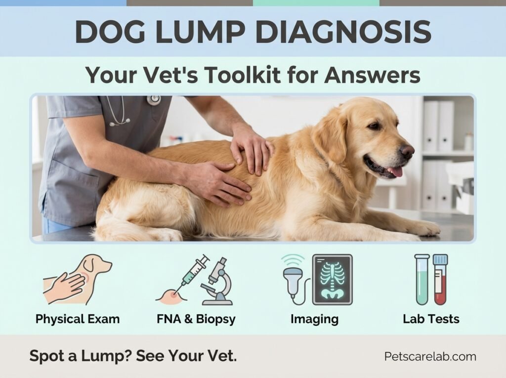
Dog Lump Diagnosis: Your Vet's Toolkit for Understanding Those Bumps
Spotting a lump or bump on your dog can send your heart racing, and that's completely normal. As pet parents, we immediately worry about the worst. The good news is, not every lump spells trouble. Many are harmless, easily treated, or might not even need attention. But some do need quick action. That's where a skilled dog lump diagnosis comes in, and our team at Petscarelab knows how important it is for you to understand what your vet is looking for.
When you bring your pup in, your veterinarian has a whole set of tools and techniques to figure out exactly what kind of lump or bump you've found.
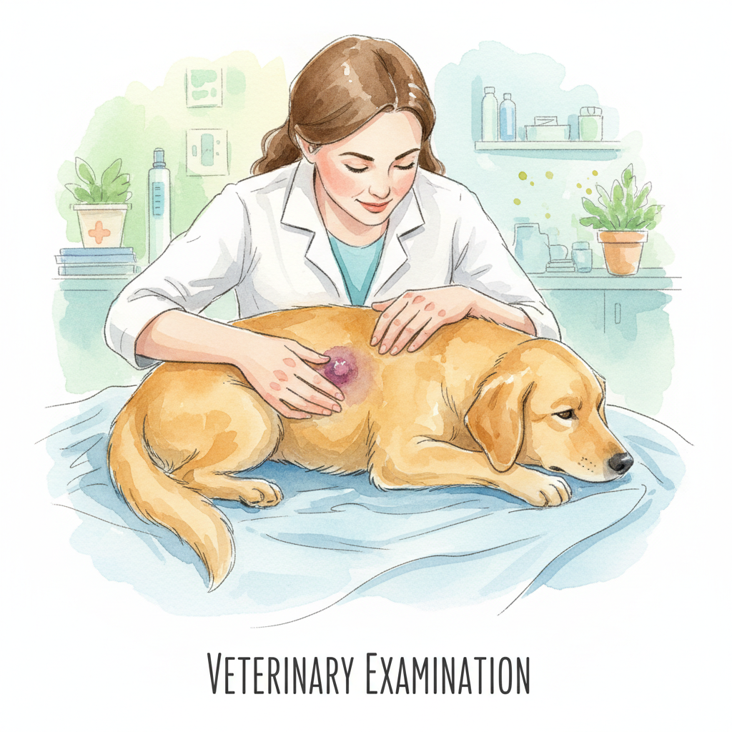
What Your Vet Sees (and Feels) First: The Physical Exam
When you first notice a lump, the first stop is always your vet's office. They'll start by giving your dog a thorough physical exam, focusing on that new growth. They'll gently feel the lump, checking its size, shape, and how it feels – is it soft like jelly, firm like rubber, or hard like a stone? Does it move easily under the skin, or is it stuck in place? And most importantly, does it hurt your dog when touched?
Your vet will also ask you some really important questions about the lump:
- When did you first spot it?
- Has it grown or changed its shape? How quickly?
- Is your dog constantly licking, scratching, or chewing at it?
- Have you noticed any other lumps or bumps on your pup before?
- Is your dog showing any other symptoms, like being unusually tired, not eating well, throwing up, or having diarrhea?
This initial chat and hands-on check-up helps your vet narrow down the possibilities and decide which diagnostic tests will give you the clearest answers.
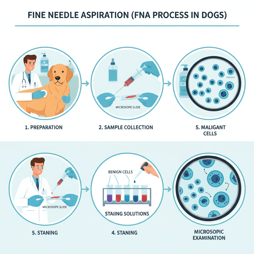
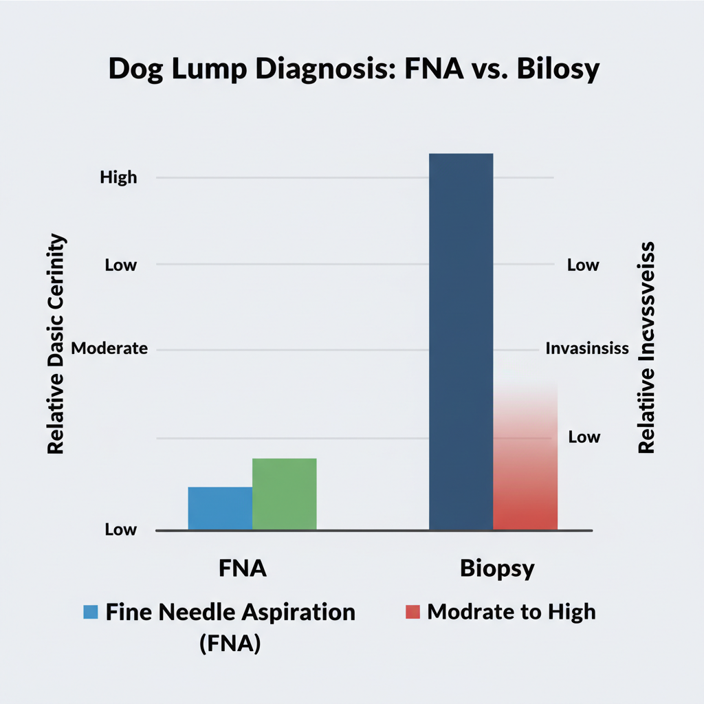
Fine Needle Aspiration (FNA): Getting a Closer Look
Often, the first test your vet will suggest for a new lump is a fine needle aspiration (FNA). It's a quick and gentle way to collect cells from the lump for a peek under the microscope.
Here’s how it usually works:
- Getting Ready: Your vet might clean the skin over the lump. Most dogs handle an FNA without needing sedation, but if the lump is in a super sensitive spot or your dog gets really anxious, they might get a mild sedative to keep them comfortable.
- Collecting the Sample: Your vet uses a very thin needle, much like the one used for vaccinations. They gently insert it into the lump and attach a syringe to draw out a tiny sample of cells. Sometimes, they'll redirect the needle a few times inside the lump to make sure they get a good, representative sample.
- Making a Smear: Those collected cells then go onto a microscope slide, where your vet spreads them out thinly to create a "smear." They're usually air-dried or fixed with heat.
- Microscopic Check: Your vet or a veterinary pathologist will then stain the slides and examine them under a microscope. They're looking for different cell types – maybe inflammatory cells, fat cells, mast cells, or even tumor cells. This helps them figure out if the lump is benign (harmless) or malignant (cancerous).
An FNA is quick, generally well-tolerated by most dogs, and often gives results fast – sometimes within minutes if your vet examines it in-house, or a few days if it goes to an outside lab. While it's incredibly helpful, sometimes an FNA doesn't give a definitive dog lump diagnosis, especially if the sample is too small or doesn't capture the true nature of the whole lump.
Biopsy: When You Need More Answers
If the FNA results aren't clear, or if your vet suspects something more serious, they might recommend a biopsy. This means taking a larger piece of tissue from the lump for a more detailed look. There are a few kinds of biopsies:
- Incisional Biopsy: Your vet removes just a part of the lump. This is often done when the lump is quite big, or its exact nature is unknown, to help plan for the best way to remove it later.
- Excisional Biopsy: With this one, your vet removes the entire lump, plus a small border of healthy tissue around it. This is often a two-in-one deal: it helps diagnose the lump and removes the problem entirely.
- Punch Biopsy: Your vet uses a special circular tool to take a small core of tissue from the lump. It's less invasive than an incisional biopsy and is often used for skin growths.
Biopsies usually need local anesthesia, sedation, or sometimes even general anesthesia, especially for excisional biopsies or if the lump is deep inside your dog's body. The tissue sample goes to a veterinary pathologist for a detailed examination, and results can take several days to a week. A biopsy offers a much more definitive dog lump diagnosis than an FNA.
It can tell you the specific type of tumor (if it's cancerous), how aggressive it might be, and, for excisional biopsies, whether it's been completely removed.
Imaging: Seeing Beyond the Surface
For lumps that are internal, deep, or if your vet is concerned the lump might be affecting structures deeper down or has spread, imaging techniques become incredibly valuable.
- X-rays: We mostly use X-rays to check out bones or to see if anything has spread to the lungs or other internal organs. If your dog's lump feels hard and stuck, an X-ray might show if it's attached to bone.
- Ultrasound: This uses sound waves to create real-time images of soft tissues. It's fantastic for looking at internal organs, figuring out if a lump is solid or filled with fluid (like a cyst), and seeing how it relates to the surrounding areas. It can also guide the needle for an FNA or biopsy of internal masses.
- CT Scan (Computed Tomography): A CT scan gives us very detailed, cross-sectional images of the body. It's super helpful for planning surgery, seeing the full extent of a tumor, and checking for spread in areas that X-rays or ultrasound can't quite reach.
- MRI (Magnetic Resonance Imaging): This offers even more detailed images of soft tissues, especially the brain and spinal cord. While not as common for general lumps, it's vital for masses that affect neurological function.
These imaging methods help your vet visualize the lump in its full context, determine its exact size and location, and look for any signs that it might have spread to other parts of your dog's body.
Blood and Urine Tests: Checking Overall Health
While blood and urine tests won't directly diagnose a lump, they give your vet important information about your dog's overall health and how their organs are functioning.
- Complete Blood Count (CBC): This can flag signs of inflammation, infection, anemia, or other systemic issues that could be connected to the lump or point to another underlying disease.
- Biochemistry Profile:
This checks how well organs like the kidneys and liver are working. It can spot metabolic changes that sometimes link to certain tumors or help your vet decide if your dog is healthy enough for anesthesia. - Urinalysis: A urine test can help detect urinary tract infections, kidney disease, or other systemic problems.
These tests are often part of a pre-surgical workup or if your dog is showing other signs of illness.
What Happens After a Diagnosis?
Once your vet has a clear dog lump diagnosis, they'll sit down with you to go over all the findings and recommend the best plan of action.
- Benign Lumps: If the lump is harmless, your vet might suggest removing it if it's bothering your dog, getting in the way of movement, or if you're concerned about how it looks. Some benign lumps, like fatty tumors (lipomas) or sebaceous cysts, can just be monitored if they aren't causing your dog any trouble.
- Malignant Lumps (Cancer): Treatment options usually include surgery (often with wider margins), chemotherapy, radiation therapy, or a combination of these. The choice depends on the type of cancer, how advanced it is, and your dog's overall health.
Finding a lump on your dog can be unsettling, but getting them to the vet quickly is crucial. With all the diagnostic tools available today, your vet can accurately identify what's going on and guide you through the best treatment plan for your beloved companion.